Dosing Calculator
With the Dosing Calculator, you’ll never have to worry about dosage errors or performing complex calculations.
Veterinary wounds – and the wounded animals themselves – are so variable that each dressing must be tailored to fit the individual case.
What follows are examples of veterinary dressings. If you do not find an example that works for you, give us a call.
A list of published articles describing dressings and treatments of animals with maggot therapy can be found at the end of this page.
At Rood and Riddle Equine Hospital, when we are applying the maggots, we try to dress the wound so that the following criteria are met:

The maggots have good access to necrotic tissue – Usually we are doing some degree of debridement prior to application of the maggots. Depending on the case this can range from a small hole in the sole of the foot to debridement of the third Phalanx. In the case of severe infections that communicate with the distal interphalangeal joint or the navicular bursa, a surgical drain can be placed and the maggots/dressing are placed at the egress of the drain. If the horse has a cast placed over the site of infection, due to instability of the hoof or distal interphalangeal joint, a small access hole is cut in the cast so the maggots can be placed and the gauze can be changed. In cases where foot casts are employed the maggots do an excellent job at keeping infection inside the cast under control.

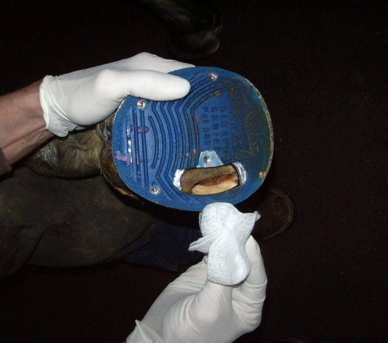
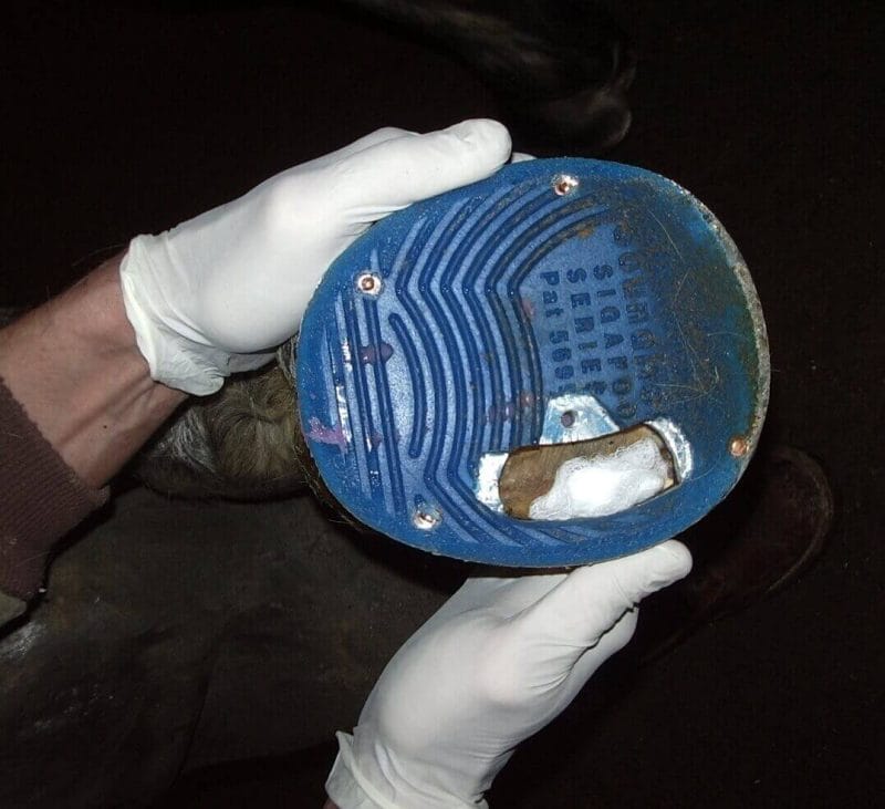
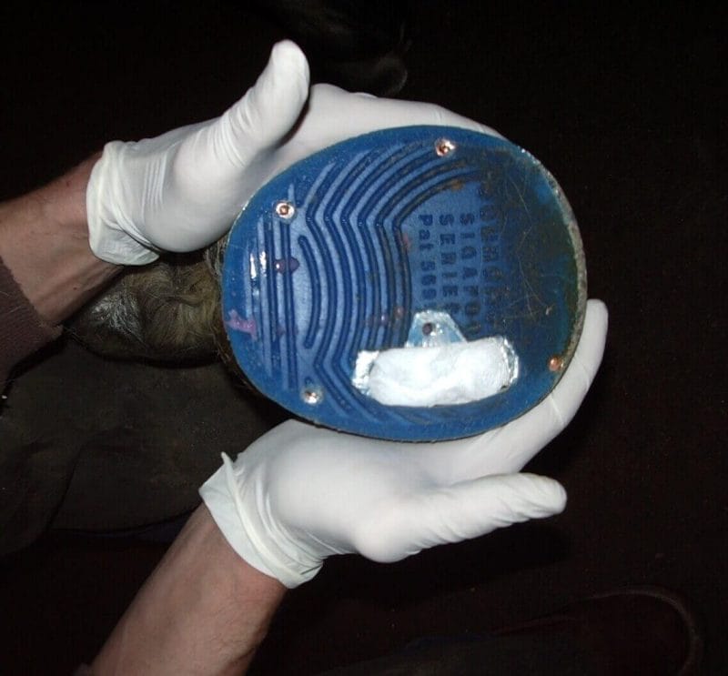
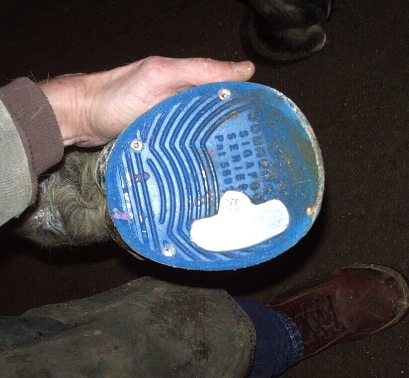
The maggots are not compressed or suffocated in the wound – Since we deal exclusively with hoof problems, we must ensure that if the maggots are placed in the bottom of the foot they are not compressed to tightly when the horse is weight bearing. Occasionally, the defect in the sole of the hoof is large enough that we can place the maggots, along with the dressing in the wound and apply a heavy foot wrap. Otherwise a shoe is applied that has a plate that offers wound protection and can be removed in order to change the gauze and monitor the progress of the maggots. We typically change the gauze and check the wound every other day. For us, the maggots mature in anywhere from 2 days to 8 days, depending on the severity of the infection.

The nature of the infection indicates the application of maggots – Cases that do not respond to maggot therapy are often infections in which proteus vulgaris is cultured from the site, especially when highly resistant strains of proteus are cultured. In these cases will often use topical disinfectants and/or systemic and regional antibiotic therapy in order to change to bacterial population in the wound prior to or in conjunction with maggot therapy.

Once a good environment has been established for the maggots they are place in the wound with the small gauze that comes with them in the vial, then cotton gauze is loosely placed over the maggots and the appropriate protection is applied. As stated above the gauze is changed every other day and maggot therapy is continued until the wound was stopped draining and looks healthy.
Berglund C: The use of maggots in canine, feline, and equine wound care. Skara 2013.
Ahmadnejad M, Rezazadeh F: Maggot Therapy for Snakebite Necrotic Wound in a Horse. Iran J Vet Surg 2021; 16(2); Serial No: 35; Pages: 156-160
Lepage OM, Doumbia A, Perron-Lepage MF, Gangl M. The use of maggot debridement therapy in 41 equids. Equine Vet J Suppl. 2012;(43):120-5.
Brevard Zoo’s Sea Turtle Healing Center doesn’t shy away from seeking out innovative science-based treatments for their patients, from raw honey to leeches. The Healing Center team recently tried a new-to-them treatment on a new patient: medicinal maggots!
With the Dosing Calculator, you’ll never have to worry about dosage errors or performing complex calculations.